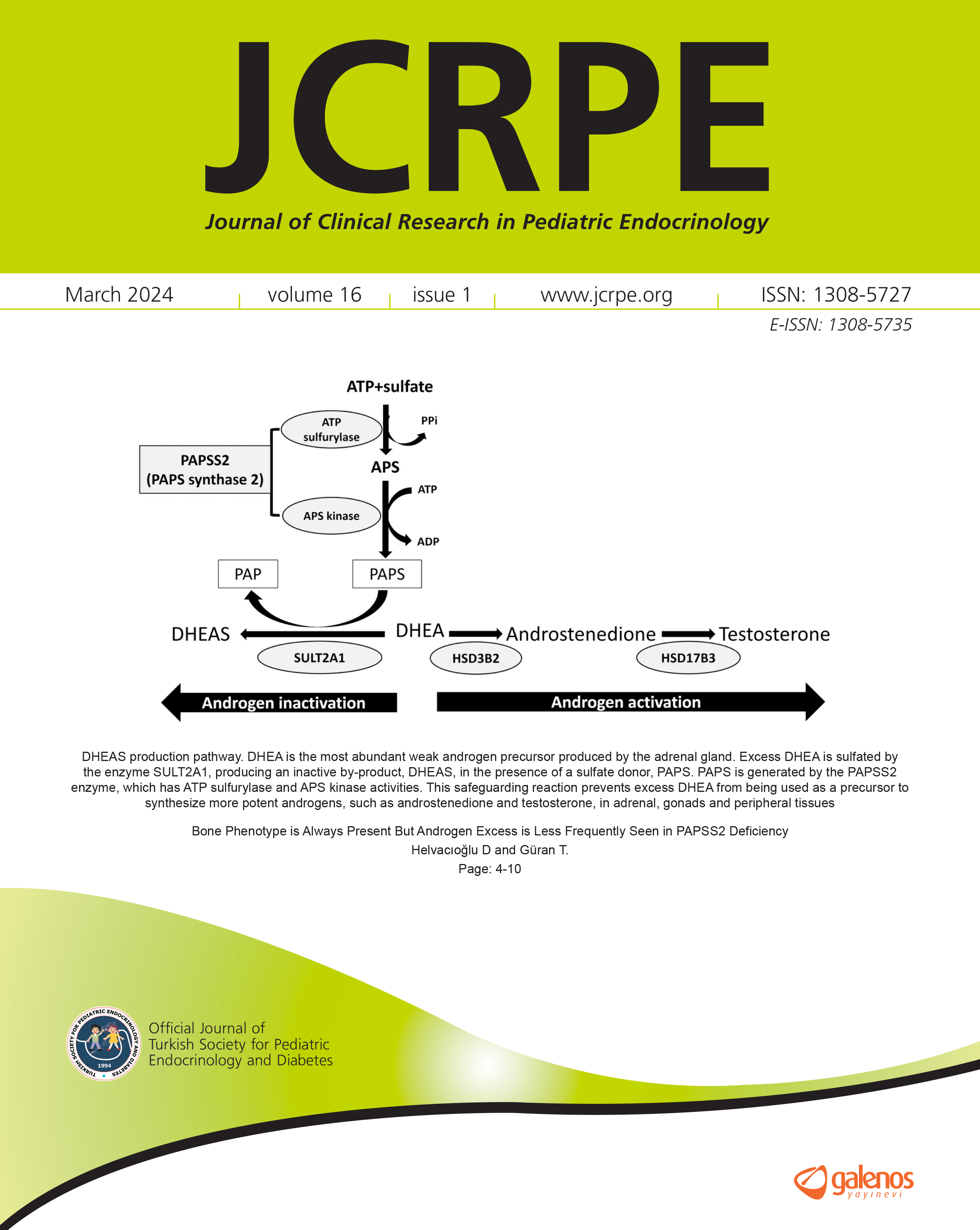


Retinal Neural and Vascular Structure in Isolated Growth Hormone Deficiency Children and Evaluation of Growth Hormone Treatment Effect
Özge Yüce1, Nuriye Gökçen Yalçýn2, Aysun Bideci1, Esra Döđer1, Hamdi Cihan Emeksiz1, Murat Hasanreisođlu2, Zeynep Aktaţ2, Orhun Çamurdan1, Peyami Cinaz11Gazi University Faculty of Medicine, Department of Pediatric Endocrinology, Ankara, Turkey2Gazi University Faculty of Medicine, Department of Ophthalmology, Ankara, Turkey
Objective: To evaluate neural and vascular retinal morphology of children with isolated growth hormone deficiency (GHD) and to determine any retinal changes due to GH treatment.
Methods: Twenty-eight children with isolated GHD and 53 age-, gender- and body mass index-matched healthy volunteers were enrolled in this prospective study. The retinal nerve fibre layer (RNFL) and macular thickness (MT) were measured, as well as intraocular pressure (IOP). The number of retinal vascular branching points were calculated. Effect of GH treatment on the retina and IOP was evaluated after one year of treatment. Measurements were also made in the control group at baseline and following the initial examination. Pre- and post-treatment changes were compared. The findings were also compared with those of the controls. The correlation between ocular dimensions and insulin-like growth factor-I (IGF-1) levels were also analysed.
Results: The number of branching points was significantly lower in GHD patients as compared with control subjects (15.11±2.67 and 19.70±3.37, respectively, p=0.05 for all comparisons). No statistically significant differences were found in mean RNFL, MT and IOP values between GHD patients and control subjects. GH treatment did not create any significant changes in the retinal vascularization or other retinal neural parameters and IOP either within the patient group or when compared with the control group. No correlations were observed between ocular dimensions and IGF-1 levels.
Conclusion: Our findings suggest that isolated GHD may lead to decreased retinal vascularization. However, retinal neural growth and differentiation were not affected by GHD. These findings may be related to the fetal development process of pituitary somatotropic cells and the retina. Additionally, GH treatment did not cause any changes in retinal neural and vascular tissues.
Manuscript Language: English